


CHẤT LƯỢNG CAO NHẤT - GIÁ THẤP NHẤT - ĐẢM BẢO SỰ HÀI LÒNG
Eye & Brain Health
20 Sản phẩm liên quan Eye & Brain Health
Eye-Brain Communication
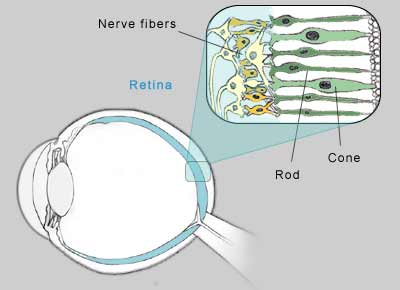 |
| Rods are responsible for night peripheral vision. Cones produce color and central vision. |
The wide, cone-shaped cells of the retina are sensitive to light. In bright environments, the chemical iodopsin increases in the cones. They become more sensitive, and we can see greater detail and color.
The long, thin rods react to lack of light. In the dark, a chemical called rhodopsin, or visual purple, increases in the rods, improving their sensitivity. Meanwhile, the cones do not receive enough light for a chemical reaction to take place, so they do not function in the dark. This provides vision in dim light, but with less detail.
The rods require about 30 minutes to be fully functional in the dark, while the cones adapt to brighter conditions in just a few minutes.
Central and Peripheral Vision
The light-sensitive rods and cones form a tightly packed network.Each eye contains 100 million rods and 3 million cones but they are not distributed evenly. Rods are mainly located in the outer edges of the retina, and cones are clustered in the center. As a result, central vision is clearest, while peripheral vision is less precise.
Vision Persistence
The image of an object on the retina is retained very briefly, fading almost instantly. But the eyes instinctively continue to gather new input. In a healthy retina, new images are received before old images fade - at the rate of about 30 times per second. This gives the appearance of an image merging into the next. It is this persistence of vision that provides the appearance of continuous, smooth movement - just as motion pictures blend one frame into another.
Binocular and Stereoscopic Vision
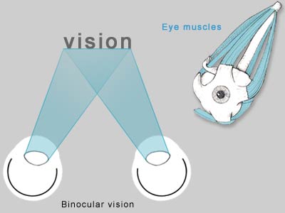 |
| Muscles coordinate the position and movement of each eye, creatin binoular vision. |
"Binocular vision" refers to sight generated from two eyes. The separate images received in each eye are combined into a single image. To see clearly, without double vision, the images must fall precisely on corresponding positions on each retina. Six muscles surrounding each eye work together to position the eyes properly so that light is focused on the center of each eye, providing clear vision. To focus on objects that are within close range, the eyes move closer together (convergence). To focus further away, the eyes move further apart (divergence). When the eyes are not aligned properly, double- vision results. Binocular vision is largely responsible for our ability to see in three dimensions. The slight difference in the angles of the images received in each eye gives images depth. This ability is called stereoscopic vision.
However, people with vision in only one eye do not necessarily see flat, two-dimensional images. Light, shade, shadows, color and relative sizes of objects contribute to depth perception.
People think and learn best in three dimensions. When scanning text quickly, we can absorb 100 letters per second - the computer equivalent of 100 bits per second. By comparison, when glancing at a three-dimensional object, we can see the equivalent of 1 billion bits per second.
Eye-Brain Connection
Visual information is received through the eyes but interpreted with the brain. Electrical signals are relayed from the retina to the brain via the optic nerve. The ability to recognize what we see lies in the occipital lobe, near the back of the head.
The Blind Spot
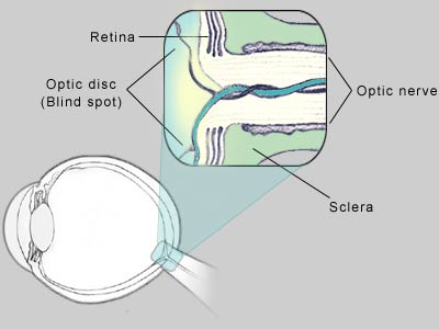 |
| The area where the optic nerve meets the retina is a "blind spot". |
In the small, round area where the optic nerve meets the retina, there are no rods or cones. If light from an object lands on this spot, the image is essentially invisible. This area is called the optic disk, or the blind spot. In effect, it's a black hole in the retina's projection screen.
This missing area in the field of vision is not normally noticeable. The brain fills this blind spot with the colors and patterns of the objects surrounding environment. It uses information received fractions of a second prior from a different distance or angle and what the opposite eye sees to create a complete, uninterrupted picture.
Everyone has this blind spot in each eye. The size of the blind spot varies from one person to another, and even from one eye to the other. To observe the effects of the blind spot, use the illustration below:
 |
| Images "disappear" when projected on the blind spot. The brain fills in the "hole" with details from the background. |
Close your left eye. Look at the dot with your right eye from about one foot away, then gradually move toward it. Be sure to keep your eye on the dot and move very slowly. At some point, the dot will disappear. That is the distance and angle at which the dot is reflected on the optic disk.
Notice that the brain fills in the blind spot. It guesses what's in the area that it can't see. In this case, it fills it with black, like the area immediately around the dot.
Similarly, if you were to draw a line across the screen and through the dot, and repeat the exercise, the dot would disappear but your brain would fill in the gap in the line.
Optical Illusions
Sometimes the brain distorts reality, incorrectly interpreting the environmental clues that surround an object. Optical illusions demonstrate this phenomenon.
Which person is tallest?
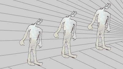 |
| Which figures are tallest and smallest? |
From an early age, people learn that the more distant an object is, the smaller it looks. In the illustration above, the person on the far right appears to be smaller than the person on the left. They are actually the same size.
People who live in the jungle often have trouble judging distances in the open country. Because their environment has not contained open, distant views, they have not learned that distant objects look smaller.
What do you see?
 |
| Do you see vase or two faces? |
Sometimes there is more than one way for the brain to interpret an image. In the illustration above, do you see a vase or two faces? The vase is white on a black background, while the faces are black on a white background.
Which square is larger?
 |
| Eye-Brain Communication Fig 6 |
In each of the two illustrations above, one square is surrounded by four rectangles larger than the center square in one drawing, smaller in the other. The center square that is surrounded by smaller rectangles appears to be larger than the center square surrounded by larger rectangles. In reality, the center squares are the same size.
The Moon
Why does the moon appear larger when it is near the horizon than it does overhead?
When the moon is close to the horizon, it is viewed along with other objects, such as mountains or skyscrapers. Without the benefit of nearby objects, the moon seems to shrink in the expansive sky. The brain misinterprets the size of the moon relative to its surroundings.
Virtual Sight
Experiments have revealed that by stimulating the retina with preprogrammed configurations of ultrasound, some blind people are able to "see" black and white outlines and images. The brain interprets the images as if light were projected on the retina.
If a camera could successfully digitize images and translate them into recognizable ultrasound impulses, it may be possible to give virtual sight to the blind.
What Your Eyes Say About Your Brain
The scientists found that people with wider veins scored worse on IQ tests in middle age. Other factors like smoking, diabetes, or socioeconomic status couldn't be to blame for the scores, says Idan Shalev, Ph.D., the study’s lead author.
What gives? Your eyes’ vessels may reflect the condition of your brain’s vessels because they're similar in size, structure, and function, says Shalev. “Eye vessels are developed from the same cells that brain vessels are developed from,” he adds.
Previous studies have linked the size of blood vessels in your eyes to risks for other diseases like dementia, cardiovascular disease, or stroke—but those studies were done in older people, says Shalev. This study found that the health of your eyes could indicate brain health at a much earlier age. The results were seen even in children.
So what does it mean for you? Pencil in the eye doctor. Even if you’re blessed with 20/20 vision, retinal imaging (a fancy term for the photo eye docs take of your eyes) does far more than test vision: It could be the easiest way yet to check in on your brain. It’s also a good way to keep track of changes if you’re at high risk for a disease like cardiovascular disease, Shalev says. Being able to compare images over time could help ID changes in midlife that hint towards problems. Otherwise, these changes could go unnoticed as they may not show symptoms until much later, he says.
Eye Disease As Marker Of Brain Health
For the study, lead author Dr Mary Haan, professor of epidemiology and biostatistics at the University of California, San Francisco (UCSF), and colleagues, used data from the Women's Health Initiative Memory Study and the Site Examination study, two sub-investigations of the Women's Health Initiative Clinical Trial of Hormone Therapy.
The findings, which they report in the 14 March online issue of Neurology, suggest that a simple eye test could look for early signs of retinopathy, and serve as a marker for cognitive changes linked to vascular disease. This would allow for earlier diagnosis and treatments that potentially reduce the progression of cognitive impairment to dementia.
Retinopathy usually results from Type II diabetes or high blood pressure (hypertension). So an early diagnosis of this eye disease could indicate early stages of these two conditions, allowing for timely changes in lifestyle or drug interventions, when they might have the most impact.
Haan told the press:
"Lots of people who are pre-diabetic or pre-hypertensive develop retinopathy."
"Early intervention might reduce the progression to full onset diabetes or hypertension," she added.
For their study, the researchers analyzed data on 511 women with an average age of 69 years at the start of a 10-year follow up during which the women underwent an annual cognition assessment that tested their short-term memory and thinking ability. One test, for example, asked them to listen to several words, and then recall them after five minutes.
They also had had an eye test in the fourth year of follow-up, and a brain scan in the eighth year.
The results showed that during the follow-up, 39 (7.6%) of the participants developed retinopathy, and on average, their scores on the cognition tests were worse than the women who did not develop the eye disease.
When they examined the brain scans, the researchers saw that the women with retinopathy had more damage in their brain blood vessels. Specifically, they had 47% more ischemic lesions or holes in the overall blood vessel structure, and 68% in the parietal lobe.
Such lesions are thought to be caused by high blood pressure. They are typically seen with vascular disease and sometimes stroke.
Another feature that was more prominent in the brain scans of the women with retinopathy was more thickening of the white matter tracks that transmit signals in the brain. These thickenings are also thought to be due to high blood pressure.
However, retinopathy was not linked to more brain atrophy, which is a hallmark of Alzheimer's disease, suggesting that retinopathy is probably not a marker for Alzheimer's disease, said Haan.
In an accompanying editorial, Dr Rebecca F. Gottesman, Assistant Professor, Neurology at Johns Hopkins Intracerebral Hemorrhage Center, writes:
"This study highlights the potential importance of retinal evaluation in understanding the extent of cerebral microvascular disease that may be present in older adults."
She points out that:
"Retinopathy was significantly associated with cognitive scores and cerebral microvascular disease independent of the effect of hypertension and diabetes," suggesting that either it "captures information beyond a simple binary yes/no diagnosis of hypertension and diabetes, or that perhaps preclinical disease is better captured with measures of retinopathy ..."
"Further studies that evaluate the role of retinal screening in individuals at risk for cognitive impairment or dementia and the impact of this screening on subsequent cognitive decline are needed," she urges.
Funds from the National Heart, Lung and Blood Institute, the US Department of Health and Human Services, Wyeth Pharmaceuticals, and the National Institute on Aging helped pay for the study.


- Giới thiệu chung
- Kết nối cùng BRIAN IR
- Chính sách
- Trợ giúp
Đối tác thanh toán nội địa

Note : Chúng tôi mang đến cho các bạn những thông tin mới nhất trên thế giới về sức khỏe và cách chăm sóc sức khỏe. Những bài viết trên duocthiennhien.com mang tính chất cung cấp thông tin, không mang tính chất chẩn đoán, hoặc điều trị.
Sức khỏe là tài sản quý của mỗi chúng ta. Khi bạn cảm thấy trong người khó chịu hoặc mệt mỏi, hãy lắng nghe cơ thể của bạn và tìm cách điều chỉnh chế độ ăn uống và tập luyện của bản thân. Nếu bạn cảm thấy chưa yên tâm về sức khỏe của mình, hãy tìm đến lời khuyên của các bác sỹ hay chuyên gia y tế có Tâm và có Tài, để nhận được những chỉ dẫn cho sức khỏe của bạn. Đừng bao giờ nản chí, và đừng bao giờ chủ quan về sức khỏe của mình. Tri thức và hiểu được ngôn ngữ bản thân mình cũng rất quan trọng, bạn nên dành thời gian để nghiên cứu thêm về sức khỏe và hiểu được nhu cầu của cơ thể mình.
Nếu bạn muốn hủy bỏ đăng ký nhận thông tin về sức khỏe từ duocthiennhien.com, xin vui lòng bấm vào đây để hủy đăng ký
Điện thoại : 0243.990.2983 -*- Hotline : 0936.27.9939
Email : hendt.brianir@gmail.com
